
Select Areas of Research
Our faculty and trainees are engaged in research in a variety of topics in medical
physics and health physics. In the section of our web site, we describe selected major
research topics by specialty and subspecialties. In medical physics, these subspecialties
include radiotherapy physics, diagnostic imaging physics, nuclear medical physics,
and medical health physics. In health physics, the subspecialties include environmental
health physics and medical health physics. For brevity, the topics were selectively
listed. Please see our listing of research publications for addition information on
recent and current research projects of our program. Publications are listed separately
for peer-reviewed journal articles and graduate theses and dissertations. These may
found elsewhere on our website.
Medical Physics Research Areas
Radiation Therapy Physics
Several faculty in the program are studying fundamental physical aspects of radiation therapy, including proton therapy, intensity modulated x-ray therapy (IMXT), volumetric modulated arc therapy (VMAT), modulated electron therapy (MET).
Additional specific details are listed below.
- Proton and Heavy Ion Radiotherapy Physics: Proton and heavy ion radiotherapy utilizes beams with a finite penetration range and sharp lateral penumbra. These allow the treatment of tumors to high doses while minimizing dose to surrounding normal tissue. Dr. Newhauser's research group investigates a variety of topics involving proton therapy, including modeling of radiation transport of primary and stray radiations, comparative effectiveness studies of ion- and photon-beam therapies, optimization of treatment planning, prediction of patient outcomes, and microdosimetry. Dr. Newhauser and his group have also researched and published on clinical aspects of proton therapy physics, including a widely read review paper on the physics of proton therapy, an AAPM Task Group Report published in 2020 and another Task Group Report accepted for publication in 2020. In recent years, research projects have included the treatment of moving tumors with heavy ion beams, including tracking of scanned beams.
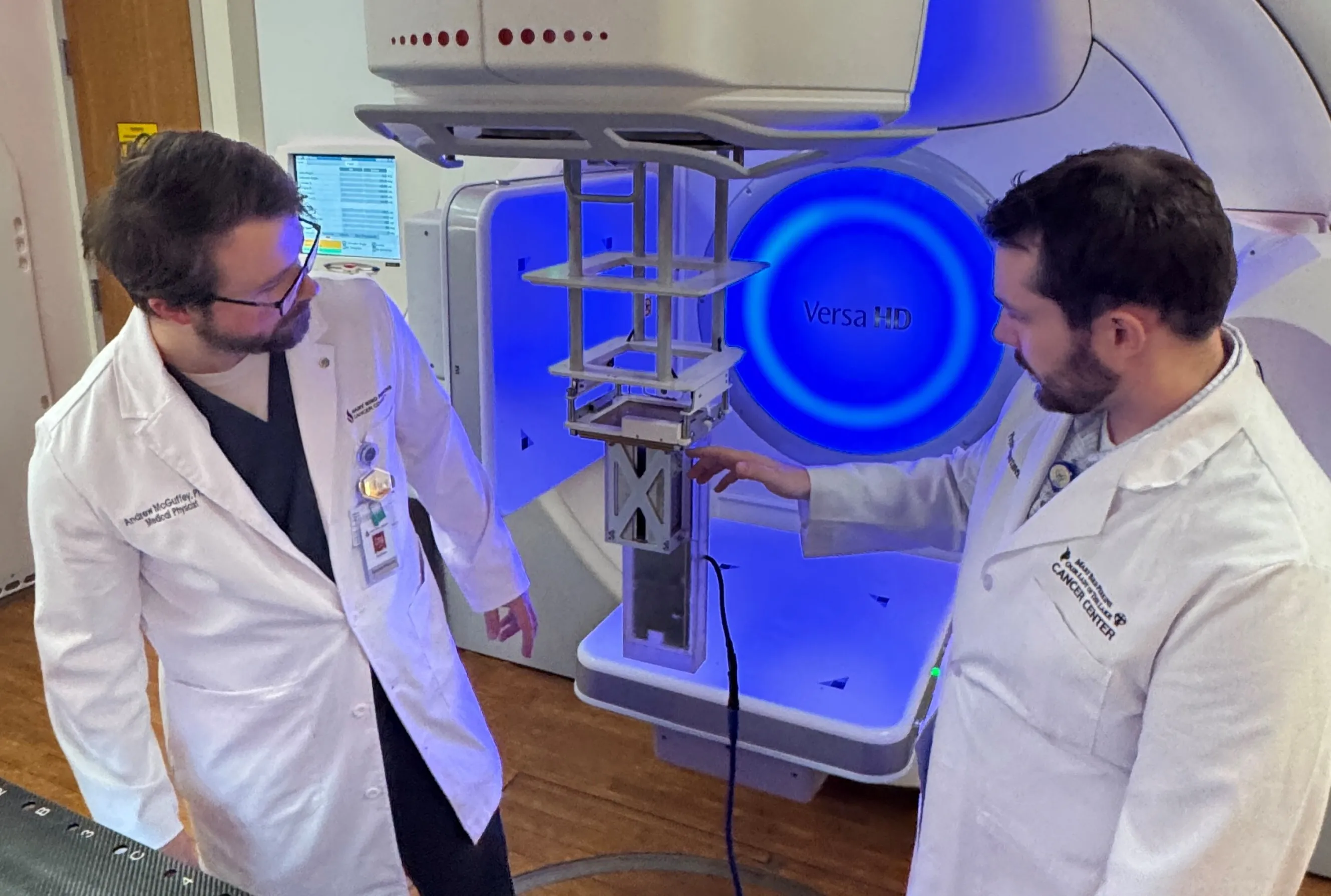 Electron Research Group (ERG): ERG aspires to discover and translate new, improved
technology for electron beam therapy of superficial patient cancers by discovering,
developing, and translating new, innovative technology for treatment planning and
delivery. Electron beams (6-20 MeV) treat cancers and other disease within 6 cm of
the skin surface, impacting as many as an estimated 50,000 USA patients annually.Members
of the research group include Drs. Pitcher and McGuffey from Mary Bird Perkins Cancer
Center, Dr. Matthews from LSU, Professor Emeritus Dr. Hogstrom, and usually two LSU
graduate students. From 2004-2025, more than $5.4 million in government and industry
grants included support for 4 PhD and 23 MS graduates, who helped publish 25 articles
in leading medical physics journals. During that span, research focused on electron
conformal therapy (ECT), the pencil beam redefinition algorithm (PBRA) for computing
patient dose distributions, and delivery devices such as dual scattering foils, applicators,
copper cutouts, electron multi-leaf collimation, and skin collimation. They also studied
Auger electron therapy using monochromatic X-rays at the LSU synchrotron radiation
facility to irradiate cells laced with iododeoxyuridine, delivering high-LET dose
to the DNA. More recently, they have focused on intensity modulated electron therapy
and developing a real-time electron energy spectrometer for accelerator beam tuning
and quality assurance.
Electron Research Group (ERG): ERG aspires to discover and translate new, improved
technology for electron beam therapy of superficial patient cancers by discovering,
developing, and translating new, innovative technology for treatment planning and
delivery. Electron beams (6-20 MeV) treat cancers and other disease within 6 cm of
the skin surface, impacting as many as an estimated 50,000 USA patients annually.Members
of the research group include Drs. Pitcher and McGuffey from Mary Bird Perkins Cancer
Center, Dr. Matthews from LSU, Professor Emeritus Dr. Hogstrom, and usually two LSU
graduate students. From 2004-2025, more than $5.4 million in government and industry
grants included support for 4 PhD and 23 MS graduates, who helped publish 25 articles
in leading medical physics journals. During that span, research focused on electron
conformal therapy (ECT), the pencil beam redefinition algorithm (PBRA) for computing
patient dose distributions, and delivery devices such as dual scattering foils, applicators,
copper cutouts, electron multi-leaf collimation, and skin collimation. They also studied
Auger electron therapy using monochromatic X-rays at the LSU synchrotron radiation
facility to irradiate cells laced with iododeoxyuridine, delivering high-LET dose
to the DNA. More recently, they have focused on intensity modulated electron therapy
and developing a real-time electron energy spectrometer for accelerator beam tuning
and quality assurance.- Image-guided Radiotherapy Physics: Drs. Fontenot, Hogstrom, Solis, and Pitcher are conducting research in image-guided radiation therapy physics, gated radiotherapy, and adaptive radiotherapy. One project concentrates on usage of orthogonal x-ray imaging using the BrainLab Novalis treatment unit for radiosurgery and radiotherapy of brain and extra-cranial cancers, e.g. spine, liver, and prostate. Another project focuses on usage of megavoltage CT scanning using the TomoTherapy HiART for radiotherapy of prostate, head and neck, and other anatomical sites.
- X-ray Capture Therapy Physics: X-ray capture therapy is a potentially new radiotherapy paradigm (chemo-irradiation) that uses monochromatic x-rays to deliver targeted radiation dose to high-Z labeled (e.g. iodine) pharmaceuticals that are preferentially taken up by cancer cells, e.g. IUdR taken up by DNA. Our research program, led by Dr. Matthews, uses the CAMD synchrotron’s monochromatic x-ray beam line to study dosimetry techniques, treatment planning dose algorithms, microdosimetry, cell biology, and small animal irradiations. A long term goal is to conduct clinical trials using a prototype laser-particle accelerator to produce monochromatic x-rays.
- Physical Aspects of Biology and Radiation Biology: Dr. Newhauser’s group is investigating novel computational models of blood flow in tissues, organs, and organisms. Blood flow is fundamental to normal physiology, disease, injury, and treatment. Dr. Matthews and colleagues are research a variety of topics in biology, including cell lysis. Dr. Dey’s research includes mathematical modeling of tumors using oncologic imaging information.
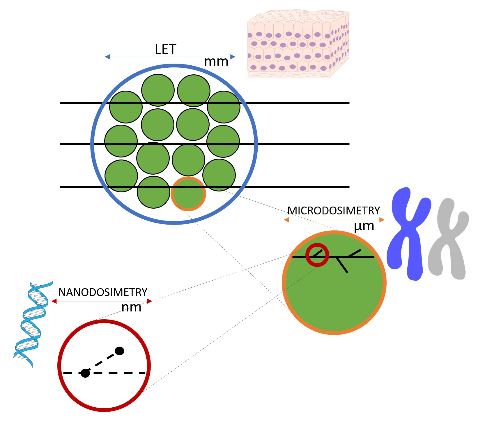
- Dr. Missiaggia's research focuses on radiation physics, microdosimetry, and biophysical modeling, with the goal of improving our understanding of how ionizing radiation interacts with biological tissues and how these interactions affect therapeutic outcomes. A central theme of her work is radiation quality, where she moves beyond traditional macroscopic descriptors to investigate how the microscopic distribution of energy deposition shapes biological damage.
Diagnostic Imaging Physics
Several of our faculty are researching advanced imaging techniques, including MRI, fMRI, CT, microCT, and multi-contrast imaging. Drs. Dey, Matthews, and Carmichael each lead research groups that are focused on diagnostic imaging projects. The areas of research include detector development, image acquisition, image reconstruction, and image analysis.
An example of a recently completed project is the development of a novel x-ray imaging
technique called endorectal digital prostate tomosynthesis (endoDPT). endoDPT is expected
to improve resolution in prostate cancer imaging for certain clinical applications.
Current efforts include applications of machine learning, quantitative testing of
image quality and patient dose on their prototype imaging system, development of tomosynthesis
image reconstruction algorithms, and development of seed localization algorithms for
low dose rate brachytherapy post-implant evaluations.
Nuclear Medicine Imaging Physics
Drs. Dey, Matthews, Carmichael and Kirby are pursuing research in medical nuclear imaging.
- Current detector development projects include high-sensitivity cardiac SPECT imaging as well as hand-held and compact CZT imaging systems for intraoperative imaging.
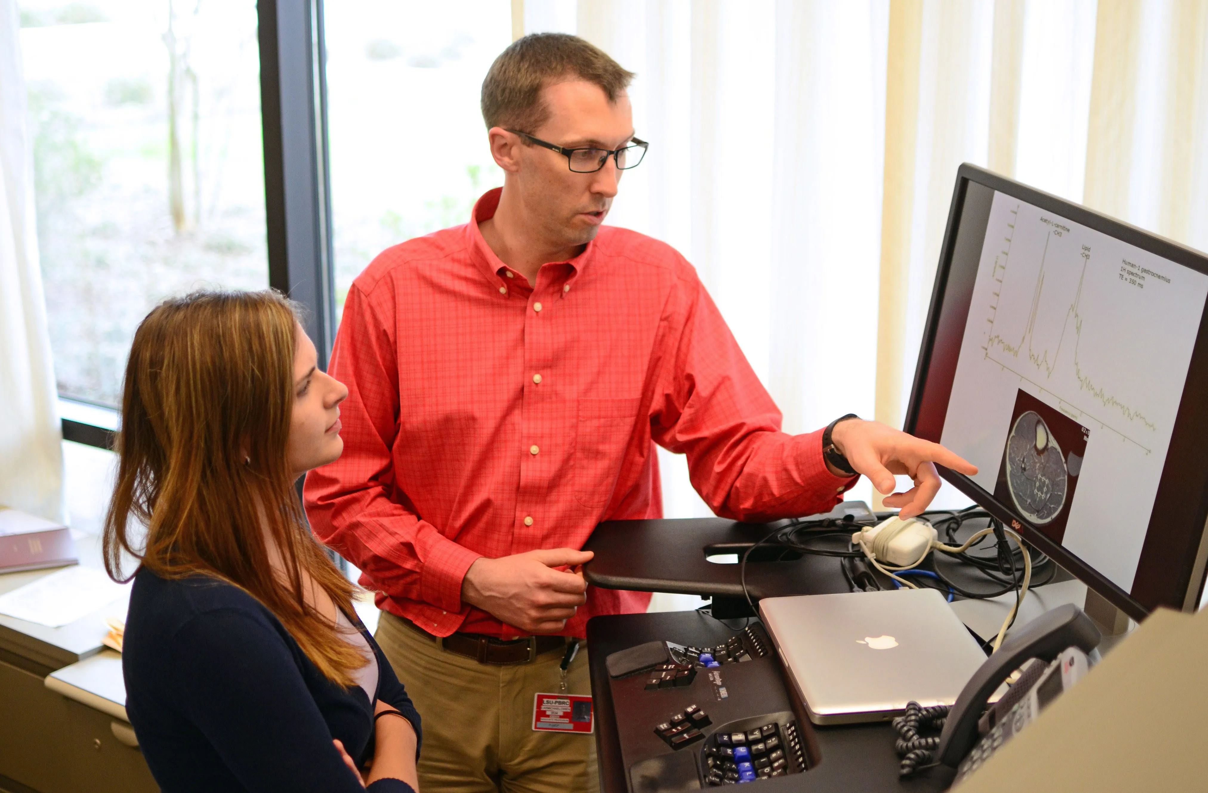 Dr. Carmichael is the Director of Biomedical Imaging at Pennington Biomedical Research
Center in Baton Rouge, Louisiana. He oversees MRI, DXA, and ultrasound data collection
and analysis at Pennington Biomedical. His laboratory works to develop new biomedical
imaging techniques that can be applied to study Alzheimer’s disease, brain aging,
metabolic disorders, and exercise, with an emphasis on the effects of lifestyle factors
on the aging brain.
Dr. Carmichael is the Director of Biomedical Imaging at Pennington Biomedical Research
Center in Baton Rouge, Louisiana. He oversees MRI, DXA, and ultrasound data collection
and analysis at Pennington Biomedical. His laboratory works to develop new biomedical
imaging techniques that can be applied to study Alzheimer’s disease, brain aging,
metabolic disorders, and exercise, with an emphasis on the effects of lifestyle factors
on the aging brain.- Graduate students with Dr. Matthews have also worked on topics such as observer performance studies for PET/CT, quality assurance methods for PET/CT, performance characterization of megavoltage CT imaging for radiotherapy applications, and dose reduction for CT lung screening.
- Dr. Dey has recently also worked on tumor modeling based on oncology imaging data.
Medical Health Physics
Drs. Newhauser, Wang, Matthews, and Missiaggia have pursued research pertaining to the protection of humans from medical radiation. This includes a variety of topics in radiation protection, including risk assessment, shielding, justification, and optimization.
Dr. Newhauser’s group is developing of a novel theoretical framework to allow the
algorithmic aggregation of risks and benefits from radiation exposures. This has
potential applications in medical, occupational, and environmental exposure settings.
Drs. Matthews and Newhauser have conducted variety of research projects on radiation
shielding of exotic radiations, including heavy ions and synchrotron radiation.
Health Physics Research Areas
Our faculty are engaged in a variety of health physics research topics that may be categorized as environmental health physics and medical health physics.
Environmental Health Physics
- Terrestrial Environmental Applications: Health physics research includes radiation
detector development with safety/security applications and intercomparisons of dosimetric
methods by Drs. Wang and Matthews. Dr. Wang and students also work on environmental
impacts of radiation use, currently including an environmental assessment of a hypothetical
low-level radioactive waste repository located in Louisiana.
Medical Health Physics
The Program’s research in this subspeciality of health physics is substantially similar
to the subspecialty of medical physics, which also goes by the same name. Please
see the description above.